
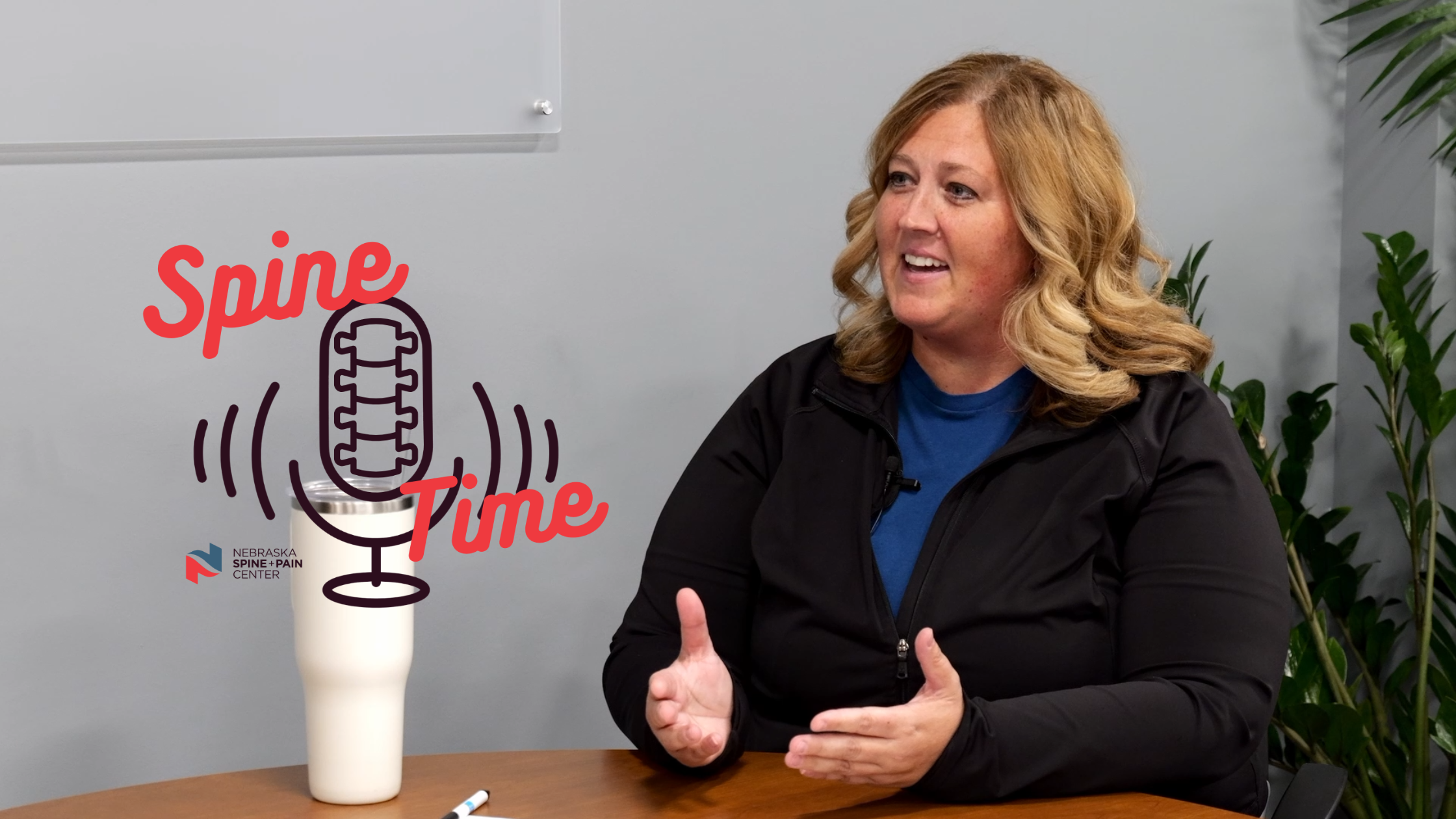
What’s actually happening inside an MRI machine? Can you take pain medication before your imaging appointment? On this episode of Spine Time, we sit down with Jen Levos, a Nebraska Spine + Pain Center MRI technologist with 20 years of experience, to break down everything patients need to know about imaging.
Tune in to learn how the right imaging test is chosen, and why having X-ray, CT, and MRI all under one roof makes it easier for patients to get answers in a single visit. You can watch the interview on our YouTube channel, or listen along on Spotify or Apple Podcasts.
Episode Transcript
Zach
Hello everybody, and welcome back to Spine Time, Nebraska Spine and Pain Center’s podcast where we discuss everything about the back, neck, and spine with Southern Nebraska’s best healthcare professionals. And today, I am lucky to be joined by Jen Levos, who is going to tell us a little bit about what you need to know about MRI, X-ray, CT scans, anything relating to that. So without further ado, I’ll just pass it over to you, Jen! Why don’t you go ahead and introduce yourself for us today?
Jen
Yeah, like you said, I’m Jen Levos. I’ve been an MRI tech for about 20 years, and I’ve been here at Nebraska Spine for about the last three.
Zach
Nice! Very excited. So today we’re covering a whole lot of topics, kind of around imaging as a whole. We do all sorts of imaging here. We do X-ray, CT scans, MRI; Why don’t you go ahead and just kind of break those down, kind of define what they are if you were an incoming patient looking to have some imaging done.
Jen
Sure! Like you said, we have all three modalities. Sometimes people forget that we do have CT and MRI in the basement. X-ray is upstairs where all the clinics are, and X-rays are a good imaging option: one, it’s fast, it’s quick, it’s a low dose of radiation, and it’s really good for bone imaging, like for fractures, arthritis. We also use it for spine alignment to show scoliosis and stuff like that. And then we have CT, which is basically an X-ray as well, but it is a more detailed imaging of the bones. And that’s typically used for hardware, pre-surgical. They do look for bone fractures and other bone abnormalities that they can look for on a CT scan. And then last we have the MRI. MRIs are great for soft tissue. It can show herniated discs, muscle, ligaments, tendons, nerve impingement, stuff like that. One good thing about MRI is it does not use radiation like CT and X-ray do. It uses a magnetic field and radio waves.
Zach
I know I’ve had X-rays done before, and I’ve seen MRIs and things done here in-house. Why do we traditionally do X-rays standing up versus a CT scan, MRI; you’re always laying down for those. What’s kind of the reason for that?
Jen
So, the reason that we perform X-rays standing up is because it helps show spine support, your body under normal conditions. If you’re laying down, you don’t always get that visualization on the X-ray. And then CT and MRI, they’re laying down; one, because they are a little bit longer. CT you’re getting thousands of images, so it’s best for you to be laying down. Same with MRI, it’s such a long exam, so the better that they’re laying down, usually they’re in a more comfortable position, and they can hold still better.
Zach
And usually how long are those tests? I know MRIs can be much longer. How much time should you expect to set aside for getting one of those done?
Jen
So X-ray alone, tops, it can take like a minute total. The X-ray itself is pretty quick, a couple seconds. CT scan, we usually tell patients it’s anywhere from two to three minutes of scanning. And then the MRI is the longest because you’re getting thousands of images. It can take anywhere from 20 to 40 minutes per exam.
Zach
We also do a thing called fluoroscopy here. I know we’ve had patients ask what that is exactly a few times. Could you kind of explain what that is? Is it, is it an X-ray?
Jen
Yes, it actually is an X-ray, but instead of being one image, it’s live imaging. A lot of times our physicians here use that for injections. What happens is in real time they can put the needle in and make sure they’re in the correct placement. Sometimes they’ll also use contrast to locate to make sure they’re exactly precise where they want to be because you want to treat that patient for pain, right? And so to be in that specific spot, they use fluoroscopy to image the area to make sure they’re triggering that point. There’s two difference contrasts for CT and X-ray is an ionized-based contrast; it shows up on X-ray. And then MRI is a gadolinium-based contrast and would show up on MRIs. CT dye does not show up on MRIs because of the metal that it’s made up of, and so that’s why there’s two different types of contrast.
Zach
I’ve heard it said many times that MRI is kind of considered the gold standard when it comes to anything relating to spine conditions when it comes to imaging. Why wouldn’t we do that every single time then if that’s going be the best thing for any condition pretty much?
Jen
Yes, MRI has been considered the gold standard. However, it’s not always the first thing that doctors should go to. It kind of depends on the patient’s history, their symptoms. Sometimes an X-ray is best for a fracture, or CT’s going be best for, say, they had surgery and they’re just looking for the hardware to make sure all that is stable. It kind of depends on what the doctor’s looking at. Yes, everyone thinks MRI is the end-all be-all, “got to get an MRI,” but that’s not always the case. It’s really dependent upon what they’re looking for and what the symptoms are and what, you know, how to help them treat the pain that they’re having. I do know that a lot of people are like, “Well, I just need an MRI.” They don’t always need that. You can see it on an X-ray. If it’s a fracture, you’d be better to visualize it on an X-ray than an MRI. So, it just kind of depends on what they’re looking at.
Zach
Earlier you were describing that MRIs use a magnetic field versus X-rays and CTs take imagery. What’s kind of happening inside those machines? How are they actually generating those images that ultimately they can be shared with the patient to show them their results?
Jen
Well, MRI is actually pretty fascinating. It uses a magnetic field that lines up tiny hydrogen atoms, which our bodies are made up of a lot of. It takes those hydrogen atoms and aligns them with a magnetic field. Then what happens is a radio wave frequency happens. A lot of times you’ll hear that loud banging, knocking noises. That’s basically knocking those hydrogen atoms out of alignment. When they come back and relax, the signal’s picked up from that, that data is then converted to the computer, and those create very detailed images of the body. And then we have CT scan, which uses an X-ray tube that spins around your body about 100 times. There’s detectors on the opposite side that measure the X-ray beam, and they see what the measurement is that crosses through the body. Then the computer combines thousands of those measurements into thin slices of the bones.
Zach
After all that data is generated and the images are kind of put together, what happens next? Once the patient comes out of the machine, what can they expect? How is that data interpreted?
Jen
We have radiologists that read the exam, and they prepare a very detailed report. That report is then given to your ordering doctor, and your ordering doctor reads that report with your underlying symptoms, the reason you came in, and kind of takes those two things and helps determine what’s the next plans for you with those two kind of things together to see what treatment’s best for you next.
Zach
I know somebody like my mother-in-law has had a fusion done before. There’s hardware in there. Is there a case where if they had to come back in for imaging again after having work done and there’s hardware now in their spine or somewhere else in their body, is there going to be any complications with imaging in that situation?
Jen
No – Actually, CTs are really good for hardware as well as MRI. The only problem you get, would be artifact. But most hardware is okay for each modality. MRI, we do a little more investigating on some sorts of metals, just because of patient safety. The biggest thing would be implants. We do a thorough history of patients before they get an MRI, just to make sure that they’re cleared to get scanned. But other than that, yes, you can get CTs, MRIs, and X-rays with hardware in your body.
Zach
Say a patient is currently dealing with pain for their condition. Is it okay for them to take any pain medication before their appointment, before they come in?
Jen
Yes. We highly recommend it, unless they are told by their provider to stop taking any medication. We can always see symptoms without pain. Sometimes that is misconceived that patients need to be in pain for us to see that on an MRI or a CT. But we do encourage you to take your pain medication, just because it does help you be comfortable. And when you’re comfortable, you hold still. And with MRIs, as we all know, they’re lengthy, they’re 20 to 40 minutes long, and they’re very sensitive to motion. So, the better you hold still, the quicker you get your pictures. So, we do encourage people to take pain medication.
Zach
On the topic of comfort, we have our new Free.Max MRI machine. Can you talk a little bit about why that’s kind of a big deal, and why it should be exciting for patients that we offer that as a machine they can have their MRIs done here?
Jen
The biggest thing with the Free.Max is it’s a larger opening. It’s 80 centimeters compared to a standard MRI, which is 60. We call it an ultra-wide bore. They have wide bores, which are 70, but this ultra-wide bore, so anybody that’s anxious or claustrophobic, the Free.Max is a great machine to have for people with those kind of symptoms – it’s easier for them to get through it. A lot of times the people are here with back pain, we’re able to bend their knees up a little bit higher to take some of that pressure off. That can help them get through the test.
Zach
Okay, to kind of wrap up today, what’s kind of the biggest, I guess most common misconception that you get about MRI in your line of work?
Jen
I think the biggest misconception is that everyone thinks MRI is the end-all be-all, everyone needs one, when in reality, all imaging modalities serve a purpose, right? It depends on what they’re looking at. X-ray is really good for looking at bone, like fractures. CT is a more detailed view of the bone, so if there was more of a bony abnormality or you had trauma from a car accident. And then MRI obviously shows great detail of soft tissue, so it really kind of depends on what they’re looking for, what the doctor’s looking for. Again, the doctor’s going to use imaging and your symptoms, your patient history, all of that combined to come up with the right treatment plan for you. The right test depends on what the answers that they’re looking for are.
Zach
Well, thank you so much for coming and talking with me today, Jen. I think it’s super cool that we offer all three imaging types under one roof. The fact that we have MRI, CT, X-ray, and they can all be potentially knocked out in one visit, I think is great for our patients. We don’t have to send them off somewhere else, and your expertise just makes it so much easier for our patients.
And thank you, our viewer or listener, for tuning in to this episode of Spine Time. You can find future and previous episodes of Spine Time on Spotify, Apple Podcasts, and our YouTube channel. Just search up Spine Time by Nebraska Spine + Pain Center to find those episodes, and we’ll see you in the next one. Bye!